Feb. 19, 2016
Tailored nanoparticles could improve cancer diagnosis and treatment

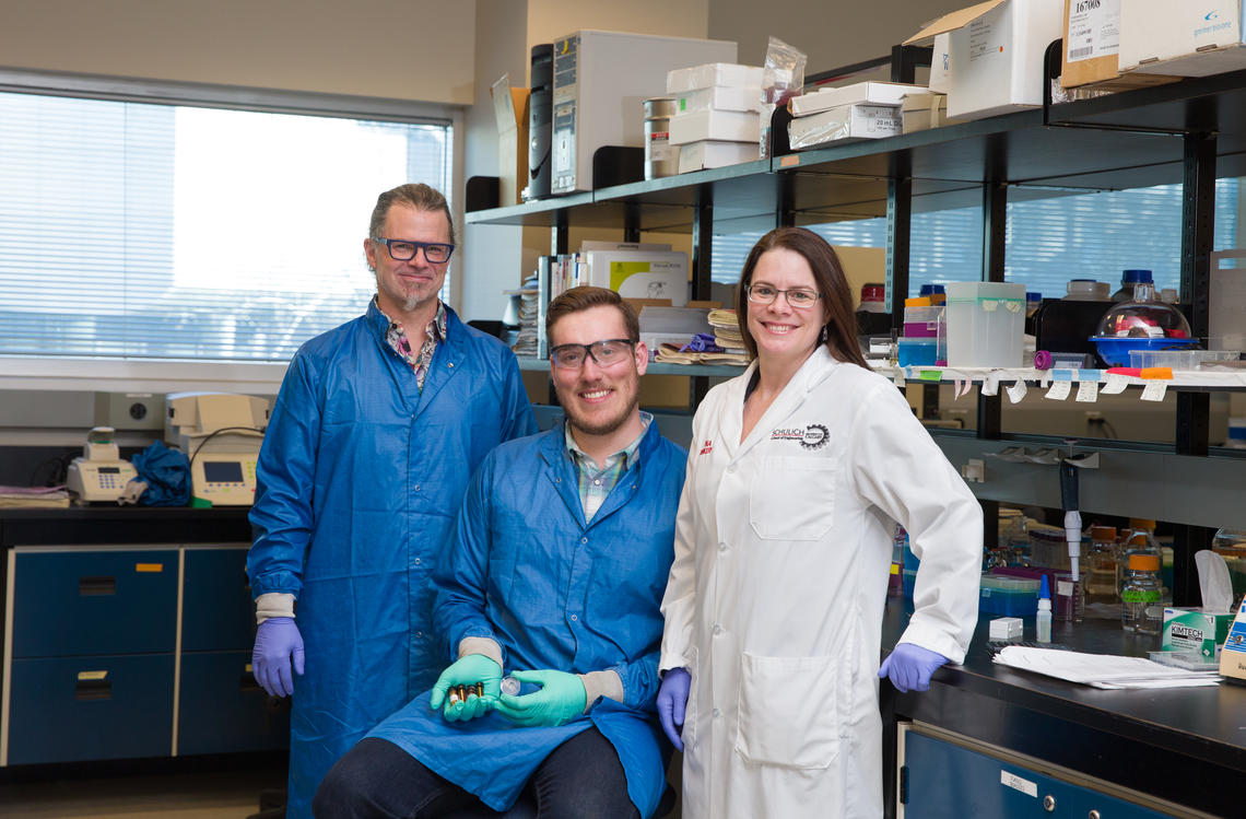
David Cramb, Christopher Sarsons and Kristina Rinker developed a novel approach to improve diagnosis
Riley Brandt, University of Calgary
Nanoparticles promise to significantly improve the diagnosis and treatment of cancer. Scientists have been designing the tiny, engineered particles (1/10,000 the width of a human hair) to carry drugs into cancer cells to destroy tumours.
However, studies show that typically less than five per cent of a nanoparticles dose delivered through the bloodstream reaches the tumour itself — greatly reducing treatment effectiveness.
Now, in a novel approach that investigated the "jello" or extracellular matrix around tumour cells, a team of University of Calgary and University of Toronto researchers has discovered why.
They found that tumour cells’ extracellular matrix — mostly collagen enveloping the tumour like jello around a bunch of a marbles — and the size of the tumour, significantly affect whether nanoparticles reach their target.
Their interdisciplinary study, a seven-year collaboration, is published this week in the Proceedings of the National Academy of Sciences of the United States of America, a globally top-ranked science journal.
“Trying to design the perfect nanoparticle hasn’t worked out as well as it should,” says David Cramb, professor and department head in the Department of Chemistry in the Faculty of Science and one of several co-authors of the study.
“Our study, which focused on tumour physiology and understanding the tumour itself, showed there’s this big barrier between the bloodstream and getting to the cells themselves — which hasn’t been looked at before.”
Paradigm shift to “personalized nanomedicine”
The research presents a paradigm shift in nanomedicine away from trying to engineer a perfect, universal nanoparticle design for cancer detection and treatment, and could lead to using specific types and sizes nanoparticles to characterize tumours and customize treatments, says the team’s study.
“Our results suggest that future clinicians will be capable of using tumour characteristics to tailor nanoparticles according to the patient — a concept called 'personalized nanomedicine.' ”
The team successfully tested this concept by using selectively sized nanoparticles to detect prostate tumours in mice, which improved targeting the tumour by more than 50 per cent.
“A tumour is not a tumour — they’re not all the same,” says study co-author Kristina Rinker, associate professor and director of the Centre for Bioengineering Research and Education in the Schulich School of Engineering.
“The cells are one part of it, but it also has this mass, this extracellular matrix that can be quite variable. And the properties of that matrix are going to affect the treatment effectiveness.”
The researchers caution that we are still many years away from physicians being able to consult a chart to see which type and size of nanoparticle to use to diagnose and treat a specific kind and volume of cancer tumour.
Graduate student built experimental system
The study’s animal model experiments, led by the University of Toronto, found that the collagen enveloping breast tumour cells vary greatly in density, both at different locations in the collagen matrix and as a function of tumour volume. This affected the nanoparticles’ ability to penetrate the collagen and reach the tumour cells.
The University of Calgary then led the building of an experimental system to study how nanoparticles diffuse into a tumour mass, penetrating the collagen and reaching the tumour cells.
Christopher Sarsons, a PhD student in Rinker’s bioengineering group and a co-author of the study, built a system that included engineered spherical gold nanoparticles, a fluid outer chamber (representing the bloodstream) and an inner chamber filled with collagen hydrogel (representing the tumour matrix). He then tracked and measured how various-sized nanoparticles moved into the gel.
The research team found — in both the mouse model and in Sarsons’ in vitro system — that getting nanoparticles to reach the tumour cells involves complex interactions; it is not just a matter of making the particles as small as possible. In some cases, depending on the tumour’s collagen density, bigger particles reached the tumor cells more effectively than smaller particles.
Complexity of cell structure not appreciated
“I don’t think the complexity of the collagen density and how it’s continuously changing has been appreciated by researchers,” Sarsons says. “We need to understand this in order to overcome this barrier and improve delivery of nanoparticles to the tumour.”
Cramb says the team’s revelations about tumour complexity could explain why many new cancer drugs that work in animal experiments subsequently fail in human trials. The findings could help improve animal models so they’re better correlated with how tumours evolve in people, he adds.
The research was funded by the Collaborative Health Research Projects, a joint venture between the Natural Sciences and Engineering Research Council and the Canadian Institutes of Health Research, with support for Sarsons from Alberta Innovates-Technology Futures and equipment funded through Canada Foundation for Innovation and Alberta Advanced Education.
The University of Calgary’s multidisciplinary Engineering Solutions for Health: Biomedical Engineering research strategy is focused on pressing health challenges like disease and injury prevention, diagnosis and treatments. These advances are also applying systems engineering principles to continuously improve the health system.
The federal government’s Research Support Fund assists Canadian postsecondary institutions and their affiliated research hospitals and institutes with the expenses associated with managing the research funded by Tri-Council agencies (CIHR, NSERC, and SSHRC). The Research Support Fund helps the university create an environment where researchers can focus on their research, collaborate with colleagues, and translate their discoveries and innovations. Read more about how UCalgary uses the Research Support Fund
